Due to its high accuracy, low maintenance requirements, and favorable physical characteristics, optical sensing technology is becoming a popular method for direct oxygen measurement across various life sciences applications, specifically in tissue, organ-on-a-chip systems, and cell culture testing.
In a study on cell hypoxia using a multi-layered model, researchers from the Oncology Department of Sidney Kimmel Comprehensive Cancer Center at Johns Hopkins University used two types of the PyroScience optical oxygen sensors in conjunction with a REDFLASH FireSting-O2 optical meter to identify hypoxic tissue regions in 3D models in vitro.
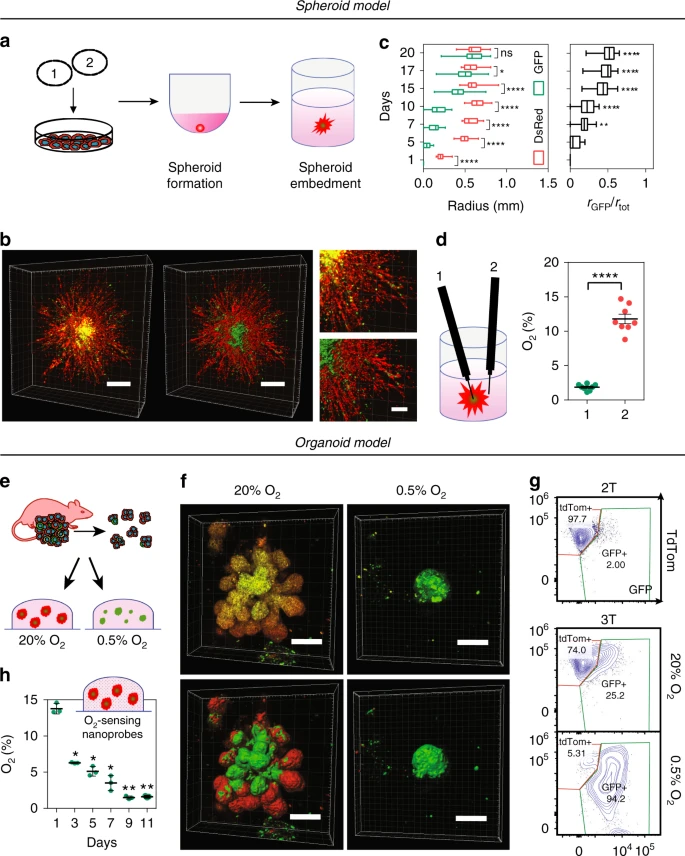
To directly measure oxygen gradients in a spheroid cultured under 20% O2 (see section d in the Spheroid model drawing below), the team used a OXR50 retractable needle oxygen microsensor (50–70 μm) . Additionally, the OXNANO oxygen nanoprobes were used to assess oxygen concentrations inside the Matrigel matrix surrounding embedded organoids (see section h in the Spheroid model drawing below). Oxygen levels were reordered every 2 days from the bottom of the well.
Additional measurement technologies and methods were used to determine other post-hypoxic cell characteristics relevant to this study. Taken together, the results revealed that:
- organoids cultured in 3D develop severe O2 gradients that range from 14% to 1.7%;
- the change in O2 levels over time may be an important environmental cue that dictates the fate of individual or clusters of cells in organoids grown in 3D cultures”.
Read the full description of this study and its results here (“Fate-mapping post-hypoxic tumor cells reveals a ROS-resistant phenotype that promotes metastasis“, Godet et al. 2020, Nature Communications).